The Importance of Using a Microsurgical Doppler in Varicocelectomy
Why intra-operative Doppler has become a key tool for protecting the testicular artery and improving outcomes in microsurgical varicocele repair.
1. Why the details of the operation matter
Microsurgical sub-inguinal varicocelectomy is already the gold-standard repair for a symptomatic or fertility-impairing varicocele—it offers the lowest recurrence (~2%) and the lowest complication rates of any technique.
But the outcome still depends on one thing the surgeon must get right dozens of times per case: telling apart the tiny veins that need to be tied off from the even tinier testicular artery that must be preserved. This is where an intra-operative micro-Doppler probe earns its place.
2. What a microsurgical Doppler actually does
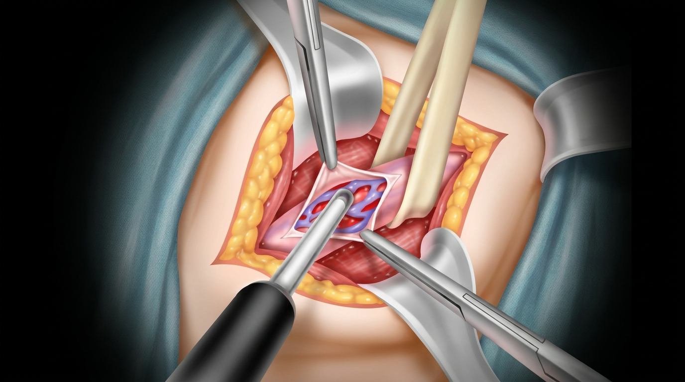
A micro-Doppler is a fine, hand-held ultrasound probe placed directly on the exposed cord structures under the operating microscope. It converts blood flow into an audible signal:
- A pulsatile signal marks an artery.
- A continuous, non-pulsatile signal marks a vein.
By listening rather than only looking, the surgeon can map the arteries before dividing any vessel—and confirm afterward that arterial flow is still intact.
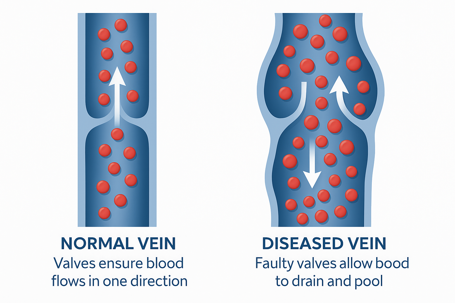
3. The core problem: arteries hide among the veins
Inside the spermatic cord the testicular artery is small (often 0.5–1.5 mm), frequently branches, and can be wrapped by dilated veins or hidden in surrounding tissue. Two things make it easy to injure:
| Challenge | Why it matters |
|---|---|
| Arterial spasm | A spasming artery stops pulsating visibly and can look like a vein |
| Multiple/accessory arteries | Missing one branch means either injury or an incomplete repair |
| Veins adherent to the artery | Blind ligation risks catching the artery in the tie |
Accidental ligation of the testicular artery can compromise blood supply and, in the worst case, contribute to testicular atrophy.
4. Microscope alone vs. microscope + Doppler
The operating microscope dramatically improved varicocelectomy by magnifying these structures. Adding a Doppler adds a second, independent confirmation:
| Step | Microscope only | Microscope + micro-Doppler |
|---|---|---|
| Finding the artery | Visual pulsation (can be lost to spasm) | Audible pulsatile signal, works even during spasm |
| Confirming a vessel is a vein | Visual judgement | Non-pulsatile signal confirmation |
| Counting all arteries | Easy to miss a small branch | Systematic sweep identifies accessory arteries |
| Checking flow after ligation | Not directly possible | Re-confirm arterial flow before closing |
5. What the evidence suggests
Studies comparing Doppler-assisted microsurgical varicocelectomy with the microscope-only approach report that intra-operative Doppler helps surgeons identify more arterial branches and preserve them, while still ligating a comparable or higher number of veins. Reported benefits include:
- More arteries identified and spared per cord, reducing the risk of inadvertent arterial injury.
- Lower recurrence in some series, attributed to more complete venous ligation once the artery is confidently mapped and set aside.
- Shorter learning curve for surgeons building microsurgical experience, because the probe gives objective feedback rather than relying on visual pulsation alone.
As with all surgical evidence, results vary by series and surgeon volume; Doppler is an aid to good microsurgical technique, not a replacement for it.
6. Practical benefits for the patient
| Benefit | How the Doppler contributes |
|---|---|
| Protected testicular blood supply | Arteries are confirmed by sound before any vein is divided |
| Lower risk of atrophy | Fewer accidental arterial injuries |
| More complete repair | Confidence to ligate all offending veins lowers recurrence |
| Fewer post-op hydroceles | Careful mapping helps spare lymphatics alongside arteries |
7. Where it fits in the operation
- Expose the cord through a small sub-inguinal incision under the microscope.
- Sweep with the Doppler to locate every arterial signal and mark it.
- Ligate the dilated veins, checking each suspicious vessel by sound before dividing it.
- Re-confirm arterial flow with the probe before closing—proof the supply is intact.
8. Key take-aways
- The single biggest technical risk in varicocelectomy is injuring the small, easily hidden testicular artery.
- A microsurgical Doppler turns an ambiguous visual judgement into an objective audible one—pulsatile means artery, continuous means vein.
- Used with the operating microscope, it helps surgeons spare more arteries, ligate veins more completely and re-confirm flow before closing.
- The result is a safer, more complete repair: lower risk of testicular injury and, in many series, lower recurrence.
- Ask your surgeon whether they perform microsurgical varicocelectomy and whether intra-operative Doppler is part of their technique.